Revolutionary method pioneered by Czech scientists reveals chromosome structure
18. 07. 2024
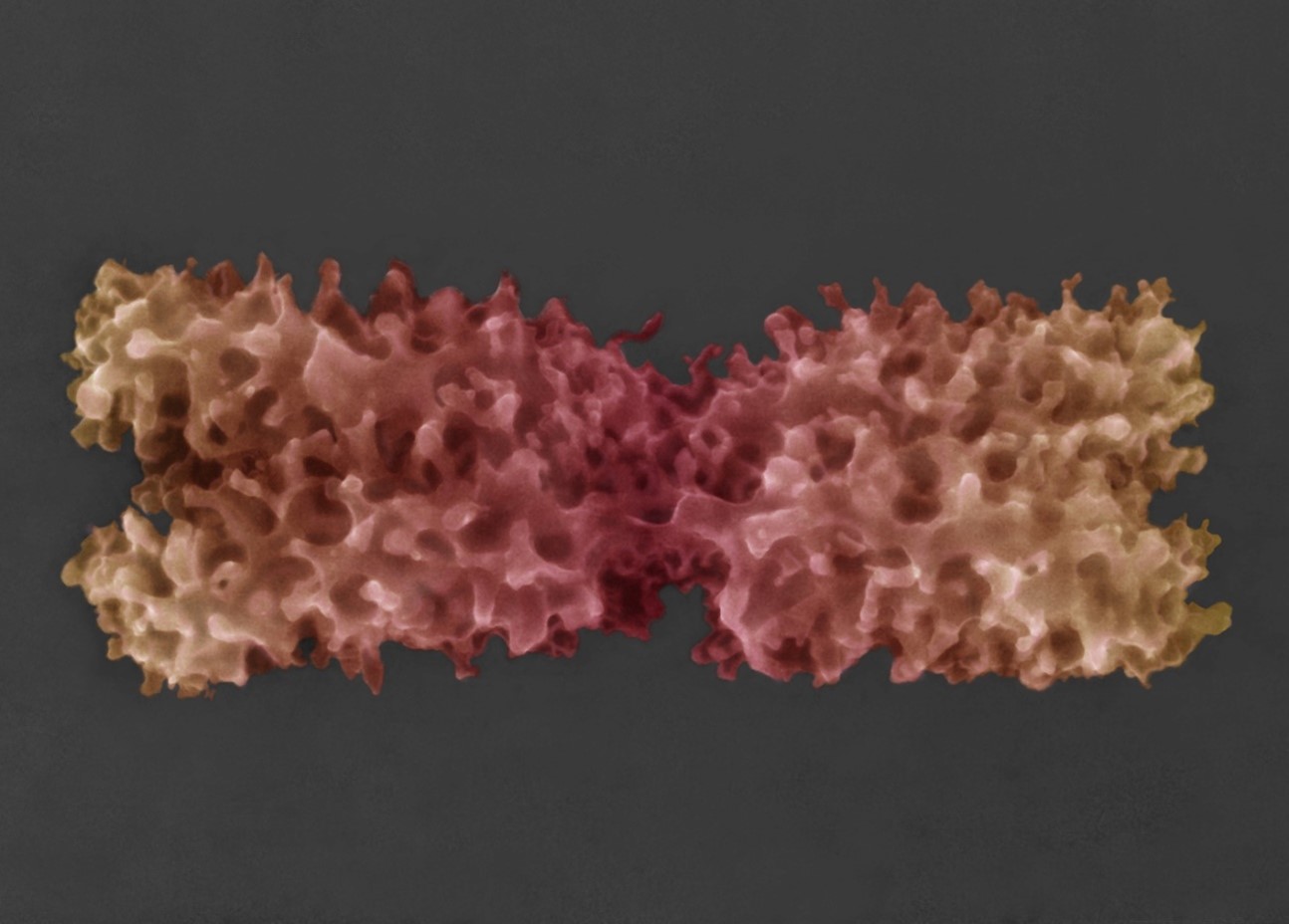
Czech scientists are the first in the world to have imaged an entire chromosome in its native state. Thanks to a new revolutionary imaging method, researchers from the Institute of Scientific Instruments of the Czech Academy of Sciences (CAS), in collaboration with the Institute of Experimental Botany of the CAS, achieved what experts from leading laboratories all over the world have been striving for. Revealing the structure of the chromosome surface, consisting of protrusions and chromatin fibers spatially organized in loops, has potential implications for fields like medicine and agriculture. The new imaging method heralds significant improvements in electron microscopy for the scientific community.
After years of research and attempts by laboratories worldwide to image biological samples at high resolution in their native state, Czech scientists have brought forth a solution. The newly developed Advanced Environmental Scanning Electron Microscopy (A–ESEM) method ushers in entirely new possibilities for studying inanimate matter, but more importantly, living matter.
Until recently, the spatial ultra-structure of biological and non-biological materials with a resolution of up to one millionth of a millimeter was imaged using Scanning Electron Microscopy (SEM), which requires samples to be observed in a high vacuum. This necessitates treatments that can damage the structure of the samples, making it impossible to observe them in their native state.
Like a car that can also drive on water and turn into a submarine
The A-ESEM imaging method makes it possible to study almost all types of living samples: plant cells and partially animal cells in their native state, small living animals, fungi, molds, mites, bacteria and protein molecules, with researchers now also preparing to study viruses. Additionally, it enables the monitoring of dynamic changes in samples due to temperature changes, drying, chemical reactions, and physical processes and stimuli.
“A conventional microscope can be compared to a luxury car, while an environmental scanning electron microscope is a luxury car that has been “tuned” and fitted with even better specifications, so it can also drive on water and turn into a submarine. It has all the functions of a classic microscope, plus many more,” explains Vilém Neděla, head of the Environmental Electron Microscopy group at the Institute of Scientific Instruments of the CAS, which developed the method.
“We use AI-based software that advises us on how to set certain imaging parameters to avoid damaging the sample. We have utilized many of our own innovations and, thanks to ultra-sensitive detectors, we observe the samples under low-dose and high gas pressure conditions and in up to 100% humidity; that is, under environmentally compatible conditions – very carefully, without inflicting any damage on them,” Neděla notes.
A–ESEM is the most versatile of all electron microscopy methods and can be used for the preparation and further physicochemical analyses of samples. According to Neděla, it is even faster, cheaper, and more suitable for studying dynamic changes in biological samples than cryo-electron microscopy, awarded the Nobel Prize in 2017. “This new method resolves the fundamental issue of the supposed incompatibility of electron microscopy with the presence of liquid water in the observed sample. That is why it is suitable for imaging living organisms and extremely sensitive nanostructures and nanosurfaces, all in high resolution,” Neděla explains.
A significant discovery for human and plant health
The potential of the new imaging method was verified by scientists in collaboration with the Institute of Experimental Botany of the CAS through the study of chromosomes – microscopic structures that store hereditary information and transfer it to daughter cells and progenies.
During cell division, chromosomes condense into microscopic, rod-like structures. For years, research teams all over the world have attempted to uncover the nanostructure of chromosomes – without success, as all available methods to date have required drastic treatment of the samples with chemicals, drying, plating, or freezing and subsequent sublimation of ice. And because the surface layer of the chromosome is extremely sensitive, it was either damaged or completely removed during such observations.
“It wasn’t until the newly developed A–ESEM method which revealed that the surface of condensed chromosomes is densely covered with numerous protrusions, loop formations of chromatin fibers averaging about 30 nanometers in size. This is the first time such spatial arrangement of the chromosome surface has been observed. Furthermore, it is highly likely that we have managed to image nucleosomes that are a mere 12 nm in size, onto which the DNA molecule is wound like a spool,” adds plant geneticist Jaroslav Doležel, head of the involved research team at the Institute of Experimental Botany of the CAS.
The results advance our understanding of the molecular nanostructure of microscopic entities – chromosomes – which transfer hereditary information from parents to offspring. In humans, chromosomal anomalies cause genetic disorders, while in agricultural crops, they lead to reduced fertility and yield. The detailed visualization of the surface structure of chromosomes will provide new insights into the structure of the genetic apparatus, enable the identification of structural defects, and contribute to the development of synthetic organisms with artificially created genetic information.
Applications of the new imaging method
The achievable resolution of A–ESEM when studying chromosomes is comparable to the resolution of scanning electron microscopy (SEM) in its vacuum conditions, which, according to Neděla, could have a significant impact on the global market for electron microscopes. “The possibility of combining A–ESEM with other imaging methods, including light microscopy, will allow researchers to image and functionally analyze not only chromosomes, but also other biological objects in their native state. The practical impact of the potential widespread adoption of the new A–ESEM method are currently beyond estimation. At the Institute of Scientific Instruments of the CAS, we are convinced that this is one of the most important discoveries ever made at our institution and is a revolutionary step forward for electron microscopy as a whole,” Neděla concludes.
The results of the years-long research conducted by the Czech scientists were published in Scientific Reports, which is part of the publishing group that includes one of the world’s most prestigious academic journals, Nature.
 Colorized image of chromosomes in their native state obtained using the newly developed Advanced Environmental Scanning Electron Microscopy (A–ESEM) method. Credit: Institute of Scientific Instruments of the CAS
Contact information:
Assoc. Prof. Ing. et Ing. Vilém Neděla, Ph.D., DSc.
Institute of Scientific Instruments of the CAS
vilem@isibrno.cz
https://www.aesemgroup.eu/
Prof. Ing. Jaroslav Doležel, DrSc., Dr.h.c.
Institute of Experimental Botany of the CAS
dolezel@ueb.cas.cz
Download the press release here.
Read also
- How lakes connect to groundwater critical for resilience to climate change
- A unique method of rare-earth recycling can strengthen the material independence
- High-energy cosmic rays dominated by heavy METALS
- Genome Tool Developed at CAS Featured in PLoS Genetics
- Why do brain cancer cells steal mitochondria?
- Secrets of the Nano- World: a new comic book about nanotechnology
- Teen duo from Slovakia and Czechia named Global Winner for clean water solution
- Professor Pavel Hozák Receives the Paul Nakane Prize
- Neutrino is lighter than previously thought
- The Earth Prize 2025 goes to Czechia and Slovakia for pioneering water purifier
Contacts for Media
Markéta Růžičková
Public Relations Manager
+420 777 970 812
Eliška Zvolánková
+420 739 535 007
Martina Spěváčková
+420 733 697 112